Warning signs of osteoporosis: Identifying the clinical indicators of progressive bone density loss
Understanding the Nature of Osteoporosis
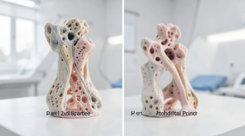
Osteoporosis is a progressive, systemic skeletal disease defined by low bone mass and the structural deterioration of bone tissue, which significantly increases bone fragility and susceptibility to fractures 9. The condition is frequently described as a silent disease because bone loss occurs incrementally over many years without noticeable physical symptoms 1. In a healthy skeletal system, bone remodeling is a balanced process where bone-resorbing osteoclasts and bone-forming osteoblasts work in equilibrium 3. When the rate of bone resorption exceeds the rate of bone formation, density decreases and the internal architecture of the bone becomes porous and weak 8.
Clinical data indicates that approximately 12.3 million Americans are currently living with osteoporosis, with an additional population segment experiencing low bone mass 9. While often associated with the aging process, it is not an inevitable consequence of getting older; rather, it is a clinical condition driven by multifactorial factors including hormonal shifts, dietary intake, and genetic predisposition 9. Because early-stage bone loss is typically asymptomatic, many individuals remain unaware of their condition until a significant mechanical failure, such as a fragility fracture, occurs 2.
Common Physical Manifestations and Structural Changes
As the disease advances, physical changes may become apparent, though they are often misinterpreted as general signs of aging. A noticeable loss of height, specifically a reduction of an inch or more, is a primary indicator that structural changes have occurred in the spinal vertebrae 3. This height loss is generally caused by the compression of the vertebral bodies, which can occur gradually or suddenly depending on the severity of the bone thinning 5. When multiple spinal fractures occur, the spine may struggle to support the weight of the body, leading to a visible stooped or hunched posture, medically referred to as kyphosis or a dowager’s hump 4.
Chronic or sudden back pain is another significant symptom that often points toward structural skeletal issues. This pain typically arises from compression fractures within the spine, where the weakened bone structure collapses under minimal stress 3. While some individuals may experience acute pain following a specific event like a sneeze or a minor lift, others may experience a gradual onset of discomfort that is frequently mistaken for other musculoskeletal conditions, such as arthritis 6. Because spinal fractures do not always cause the level of pain expected from a broken bone, they often remain undiagnosed until a physical examination reveals a change in height or posture 6.
The Significance of Fragility Fractures
A fragility fracture, defined as a break occurring from a fall at standing height or less, is often the first clinical indicator that an individual has reached an advanced stage of osteoporosis 4. These fractures are distinct from high-impact injuries because they occur despite minimal trauma, such as stumbling, picking up a grocery bag, or simply moving in a way that would not affect a healthy, dense bone 4. While any bone can be affected, clinical research highlights the hip, wrist, and spine as the most common sites for these fractures 1. The economic impact of these fractures is substantial, with the cost of managing osteoporosis-related care rising significantly as the population ages 9.
| Common Fracture Site | Typical Mechanism |
|---|---|
| Wrist | Reflexive impact during a minor fall or stumble |
| Spine (Vertebrae) | Compression from daily movement or weight-bearing |
| Hip | Minor fall or sudden shift in weight while standing |
| Ribs/Pelvis | Rare, often triggered by minor trauma or coughing |
Subtle Markers and Risk Indicators
Beyond the skeletal system, researchers have identified several subtle markers that may correlate with lower bone mineral density. Receding gums can serve as a potential indicator of diminished jawbone density, which is often linked to the systemic loss of bone mineral density elsewhere in the skeleton 3. Furthermore, clinical studies have suggested that reduced grip strength is a measurable clinical marker associated with decreased bone mineral density and an elevated risk for future osteoporotic fractures 3. While these indicators are not diagnostic, they are increasingly recognized in clinical settings as signs that warrant further assessment of bone health.
Changes in fingernail health, such as increased brittleness or weakness, have also been investigated as potential, though non-specific, indicators of broader skeletal health issues 3. These signs should not be viewed as conclusive proof of osteoporosis, but when they appear in individuals with established risk factors, they serve as useful prompts for a professional medical consultation. It remains critical to distinguish between these soft markers and the definitive diagnostic threshold provided by medical imaging technologies, which provide the only reliable way to measure true bone density 7.
Diagnostic Standards for Bone Density
The definitive method for identifying osteoporosis before a fracture occurs is through standardized bone density testing, with the gold standard being dual energy X-ray absorptiometry (DEXA) 7. This diagnostic tool uses low-dose X-rays to calculate bone density at critical sites, including the lumbar spine and the hip, providing clinicians with a T-score that indicates how much the patient’s bone density deviates from that of a healthy young adult 7. The procedure is non-invasive, quick to perform, and requires significantly less radiation than a standard chest X-ray, making it a safe primary screening tool for those at elevated risk 7.
Physicians typically recommend screening based on specific criteria, such as reaching age 65, having a history of early menopause, prolonged use of corticosteroid medications, or a documented family history of osteoporosis 1. In some cases, ultrasound technology may also be utilized to estimate bone mineral density in peripheral areas, though it is generally viewed as a secondary measurement compared to DEXA 7. Regardless of the modality, early screening is the only effective strategy for identifying low bone mass (osteopenia) before it transitions into clinical osteoporosis, allowing for early intervention and risk management 3.
Clinical Evaluation and Risk Management
When an individual presents with symptoms such as height loss, persistent back pain, or a fragility fracture, clinical evaluation involves a multi-disciplinary approach. Specialists, including endocrinologists, rheumatologists, and orthopaedists, often coordinate to manage patients diagnosed with the condition 3. The primary objectives in clinical management are to stabilize bone density, minimize the risk of subsequent fractures, and maintain physical mobility for as long as possible 3. Patients are encouraged to discuss their personal and family health histories with their primary care provider to determine an appropriate timeline for screening and preventative measures 1.
Ongoing maintenance realities for those diagnosed with osteoporosis often include lifestyle modifications, targeted physical activity to support bone strength, and, when indicated, pharmacological treatments designed to inhibit bone resorption or stimulate bone formation 8. Regulatory oversight of these treatments ensures that therapy is tailored to the specific needs of the patient, balancing the efficacy of bone-strengthening medication with the potential side effects of long-term use. By engaging with health professionals proactively, individuals can significantly impact the trajectory of their bone health and mitigate the potential for severe skeletal decline 5.
Sources
- Mayo Clinic: Osteoporosis Symptoms and Causes
- Cleveland Clinic: Understanding Osteoporosis and Risk Factors
- Penn Medicine: Clinical Overview of Osteoporosis
- NHS Inform: Osteoporosis and Fragility Fractures
- GoodRx: Early Signs and Detection of Bone Density Loss
- Royal Osteoporosis Society: Symptoms and Spinal Impact
- Harvard Health: Diagnostic Imaging and DEXA Scans
- Acibadem Hospitals Group: Risk Factors and Clinical Screening
- Medscape: Diagnosis and Management of Progressive Bone Disease
Authored by 24Trendz team