Symptoms of Raynaud's disease in toes: Clinical Analysis of Presentation and Vascular Response
Understanding the Clinical Presentation of Raynaud's Disease in the Toes
Raynaud's disease, or Raynaud phenomenon, is a disorder characterized by the intermittent narrowing of small blood vessels known as digital arteries 4. When these vessels constrict in response to cold temperatures or emotional stress, blood flow to the extremities, particularly the fingers and toes, is temporarily reduced 1, 7. This physiological overreaction is a classic hallmark of the condition, leading to observable changes in skin tone and sensory perception that can last from a few minutes to several hours, depending on the severity of the trigger and the speed of rewarming 2, 5.
While many individuals associate these vascular episodes primarily with the hands, toe involvement is equally common and can be equally disruptive to daily life 4, 5. The condition is categorized into two forms: primary, which occurs independently, and secondary, which is associated with underlying systemic conditions such as autoimmune diseases 1, 3. Recognizing the specific sensory and visual symptoms in the feet is critical for distinguishing Raynaud's from other circulatory issues or minor cold-weather discomfort 2.
The Triphasic Color Change Mechanism
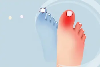
A primary indicator of a Raynaud's attack in the toes is a distinct, sequence-based color transformation of the skin 2. Initially, the toes may appear pale or white as the vasospasm occludes the flow of arterial blood to the area 3, 4. As the oxygen supply is depleted, the skin often takes on a blue or purple hue, a state known as cyanosis 2, 4. This transition represents the physiological struggle of the tissues to maintain function despite restricted circulation 2.
The final phase occurs when the vascular spasm releases, allowing oxygenated blood to rush back into the digits, a process called reactive hyperemia 3, 4. During this stage, the toes often flush a bright red color 2, 7. While this triphasic sequence-white to blue to red-is considered the classic clinical presentation, not every patient experiences all three colors in that exact order 2, 3. The marked nature of these changes is a significant diagnostic clue, separating the condition from normal cold-weather reactions 2.
Sensory Symptoms and Patient Experience
Beyond visual changes, patients with Raynaud's disease in their toes frequently report specific sensory alterations that accompany the vascular crisis 5. During the initial ischemic phase, when blood flow is blocked, toes often feel excessively cold and may experience numbness or a loss of tactile sensation 1, 4. As the circulation returns during the reperfusion phase, the sensory experience often shifts dramatically, leading to sensations of throbbing, tingling, or a sharp burning pain 2, 4.
These symptoms are often described as a pins and needles sensation by many patients as they begin to rewarm their feet after exposure to drafty environments or cold air 5. The intensity of this pain varies significantly between individuals, with some finding it manageable while others experience it as a severe, acute discomfort that disrupts physical activity 1. The emotional impact of anticipating these painful flare-ups in cold climates, such as those found in the Midwest, is a common concern for those managing chronic Raynaud's 5.
Distinguishing Between Primary and Secondary Raynaud's
Clinical distinction between primary and secondary Raynaud's is fundamental to determining the necessary level of care 3. Primary Raynaud's is considered benign, typically appearing between the teen years and age 30, and is not associated with long-term tissue damage 3. In contrast, secondary Raynaud's often presents after age 30 and is linked to systemic health issues, including lupus, scleroderma, or Sjögren's syndrome 3. Secondary forms carry a higher risk of complications and require more comprehensive medical monitoring 1, 3.
Healthcare providers often use specific markers to differentiate these types, such as nailfold capillaroscopy and autoimmune blood tests 3, 9. Normal capillary patterns are usually found in the primary form, whereas abnormal patterns like giant loops or avascular areas suggest a potential secondary etiology 3, 9. A positive result on antinuclear antibody (ANA) tests, which is often associated with connective tissue diseases, can also serve as a diagnostic indicator for the secondary type, which carries an association with higher long-term risks 3, 9.
Complications and Warning Signs
While primary Raynaud's is rarely associated with permanent tissue death, severe or secondary cases can lead to chronic complications 1, 3. Recurrent, prolonged episodes of ischemia can result in dry, cracking, or splitting skin on the toes 2. In more extreme scenarios, the lack of consistent blood flow may lead to the formation of painful digital sores or ulcerations, and in the most severe instances, gangrene can occur due to tissue death 2, 3. These developments represent a critical escalation in the disease's clinical profile.
Individuals should seek medical evaluation if they notice symptoms that persist or become progressively worse, particularly if they develop ulcers, pitting, or sores that fail to heal 3, 8. Additionally, the onset of symptoms for the first time after age 30, or the appearance of clinical features on only one foot, are considered significant warning signs that warrant professional clinical investigation to rule out underlying systemic or peripheral vascular conditions 8.
Current Approaches to Management
Management strategies for Raynaud's focus on preventing the vasospasm trigger and reducing the frequency of attacks 1. The first-line approach is consistently centered on conservative lifestyle modifications, such as cold avoidance and the use of layered, insulating socks or heated insoles to maintain digital temperature 3. Research indicates that these behavioral adaptations can reduce the frequency of attacks by 40% to 60% in many patients 3. Smoking cessation is also universally recommended, as nicotine can contribute to vascular constriction 3.
For cases where lifestyle modifications are insufficient, particularly in secondary Raynaud's or moderate-to-severe primary presentations, pharmacological interventions may be considered 3. Calcium channel blockers, such as nifedipine or amlodipine, are frequently utilized to promote vasodilation of the peripheral arterioles, which can significantly reduce both the frequency and severity of vascular attacks 3. The evidence-based hierarchy of care prioritizes these conservative methods before escalating to more intensive clinical treatments 4.
Sources
- Yale Medicine, Raynaud Phenomenon Fact Sheet
- Yale Medicine, Raynaud Syndrome: It’s Not Just Cold Fingers and Toes
- Michigan Foot Doctors, Raynauds Phenomenon in Feet: Causes Treatment 2026
- Michigan Foot Doctors, Raynauds Phenomenon in the Feet: Cold-Induced Vasospasm
- City Step Podiatry, Raynaud’s in the Feet: Symptoms and Triggers
- Radiopaedia, Raynaud Phenomenon Reference Article
- MAHESA Malahayati Health Student Journal, Primary Raynaud’s Disease in a Young Adult: A Case Report
- The Dudley Group NHS Foundation Trust, Peripheral Ischaemia Patient Information
- Medscape, Abnormal Nailfold Capillaries and Autoantibodies Linked to Higher Mortality in Raynaud’s
- European Medical Journal, Anti-Neutrophil Cytoplasmic Antibody-Associated Vasculitis Case Report
Authored by 24Trendz team