Warning signs of pulmonary embolism: Clinical identification and emergency protocols
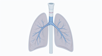
A pulmonary embolism (PE) is a critical, frequently underdiagnosed medical condition occurring when one or more pulmonary arteries in the lungs are blocked by a blood clot 17. This blockage effectively reduces or halts blood flow to sections of the lung tissue, preventing adequate oxygenation of the blood 1. Because this condition is often fatal if left untreated, recognizing the physiological markers and clinical indicators is paramount for seeking the appropriate level of care 12.
Understanding the Pathophysiology of Pulmonary Embolism
The majority of pulmonary embolisms originate as blood clots in the deep veins of the legs, pelvis, or arms, a condition formally identified as deep vein thrombosis (DVT) 27. When a portion of this thrombus breaks free, it becomes an embolus and travels through the circulatory system until it reaches the narrower arteries of the lungs, where it becomes lodged and obstructs blood passage 24. The severity of the event is largely determined by the size of the clot, the volume of the lung affected, and the existing cardiovascular health of the individual 39.
While pulmonary embolism is statistically linked to high mortality rates, early diagnosis significantly improves outcomes 27. Clinicians categorize the risk based on the specific impact the blockage has on the heart and lungs, which guides the subsequent treatment path, ranging from specialized medications to surgical intervention 19. Because symptoms can mimic other respiratory or cardiac issues, medical evaluation is required whenever sudden changes in breathing or chest comfort occur 38.
Common Respiratory and Cardiac Warning Signs
The most frequently reported indicator of a pulmonary embolism is the sudden onset of unexplained shortness of breath, which may occur at rest or during physical exertion 18. This is often accompanied by sharp, localized chest pain that typically worsens when the patient attempts to take a deep breath or cough 13. This sensation is sometimes described as similar to a heart attack, leading to confusion regarding the primary cause of distress 310.
Physiological responses to the reduced blood flow through the lungs often include a noticeably rapid or irregular heart rate 15. Patients may also experience a persistent cough that occasionally produces mucus streaked with blood 14. Such symptoms reflect the acute strain placed on the heart as it attempts to circulate blood through the compromised pulmonary system, potentially leading to a drop in overall systemic blood pressure 36.
Systemic Indicators of Hemodynamic Instability
When a pulmonary embolism leads to severe hemodynamic instability, the body may exhibit signs of systemic shock 16. Clinical presentations include lightheadedness, dizziness, and sudden episodes of fainting, known medically as syncope 13. These occurrences are critical indicators that the brain and other vital organs are not receiving adequate blood flow, necessitating immediate emergency assessment and medical stabilization 38.
Physical observation may reveal further signs of distress, such as excessive sweating or clammy, cool skin 17. In instances of profound oxygen deprivation, a patient may exhibit cyanosis, which is a bluish discoloration of the lips or nail beds 39. These signs, combined with general confusion or severe weakness, serve as urgent markers that the pulmonary blockage has reached a state of critical impact on the patient's circulatory and respiratory stability 18.
Recognizing Deep Vein Thrombosis (DVT)
Because the precursor to most pulmonary embolisms is DVT, observing the limbs can provide vital early warning signs 29. DVT typically presents as swelling in one leg, often accompanied by localized pain or tenderness that some individuals compare to a muscle cramp or charley horse 35. The affected area may also feel warm to the touch or show visible redness and skin discoloration, signaling a potential clot that remains in the peripheral veins before it potentially migrates to the lungs 35.
| Indicator | Clinical Characteristic |
|---|---|
| Shortness of Breath | Sudden onset, worsens with activity |
| Chest Pain | Sharp, worsens with deep breathing |
| Cardiac Signs | Rapid, irregular heartbeat |
| Leg Symptoms | Unilateral swelling, redness, tenderness |
| Systemic Signs | Lightheadedness, syncope, sweating |
Heightened vigilance is recommended for those who have recently experienced extended periods of immobility, such as long-distance travel, surgical recovery, or prolonged bed rest 25. Identifying these peripheral signs early can lead to diagnostic imaging or blood tests, such as a D-dimer test, which helps healthcare providers confirm the presence of clotting before a full pulmonary event occurs 39.
Diagnostic Processes and Clinical Assessment
Diagnostic protocols for suspected pulmonary embolism are designed to distinguish the condition from other lung or cardiac illnesses that present with similar symptoms, such as pneumonia or anxiety 38. Healthcare providers typically perform a physical examination that includes listening to the heart and lungs, checking blood pressure, and inspecting the legs for signs of DVT 35. Medical history is also reviewed to account for risk factors like active cancer, family history, or use of hormone therapy 14.
Imaging studies are essential for confirming the location and extent of the blockage. Computed Tomographic Angiography (CT PA) is frequently used as it provides detailed analysis of the pulmonary vessels via contrast dye 39. Additionally, an electrocardiogram may be conducted to record heart activity and identify signs of strain caused by the blockage, helping to rule out alternative cardiovascular events 35.
Risk Factors and Preventive Considerations
Pulmonary embolism risk is elevated by various life situations and health conditions that increase blood viscosity or restrict flow. Major risk factors include active malignancy, genetic or acquired clotting disorders, and chronic conditions that lead to prolonged immobility 14. Furthermore, major orthopedic surgeries, severe injuries, fractures, and pregnancy are recognized clinical triggers that necessitate professional monitoring for signs of thrombosis 49.
Effective prevention and management strategies center on maintaining circulation and early detection. Medical guidelines emphasize the importance of movement, proper hydration, and adherence to prescribed prophylactic therapy for patients identified as high-risk 9. Because even mild symptoms like fatigue or discomfort with breathing can mask the early stages of a pulmonary blockage, professionals emphasize the need for immediate clinical review when any of the identified warning signs present suddenly 810.
Sources
- American Heart Association: Pulmonary Embolism
- Cleveland Clinic: Pulmonary Embolism (PE)
- American Lung Association: Symptoms and Diagnosis
- Penn Medicine: Pulmonary Embolism Symptoms and Causes
- Stony Brook Medicine: Symptoms, Risks and Prevention Guide
- American Heart Association: New Guideline on PE
- AIA: Five Warning Signs of PE
- Acibadem Hospitals Group: Symptoms and Next Steps
- Endeavor Health: Signs and Prevention
- Acibadem Hospitals Group: Signs and Emergency Care
Authored by 24Trendz team