Home recovery for Achilles tendon repair: A clinical guide to timelines and safety
Home recovery for Achilles tendon repair begins with protecting the surgical repair, controlling swelling, and following the weight-bearing instructions provided by the treating surgical team. Early functional rehabilitation is increasingly used because controlled weight-bearing and ankle movement can support faster recovery without increasing adverse events when appropriately supervised. 1
What to expect during the first two weeks
During the first postoperative phase, the ankle is commonly held in a splint or cast with the foot pointed downward, a position intended to reduce tension on the repaired tendon. Non-weight-bearing mobility generally requires crutches or a knee scooter. Elevation and icing are used to manage pain and edema, while the incision remains protected during initial healing. 2
A follow-up appointment often occurs about 10 to 14 days after surgery. At that stage, sutures may be removed and the patient may transition into a controlled ankle-motion boot with heel lifts. Some protocols begin weight-bearing as tolerated in the boot and gentle active motion, but the surgeon's instructions take priority because repair technique, tissue quality, and individual healing affect progression. 3
Immobilization and weight-bearing progression
Recovery protocols commonly use several weeks of protection before normal walking resumes. Research information describes an initial non-weight-bearing period followed by protected weight-bearing in a boot, with some protocols extending non-weight-bearing for 6 to 12 weeks. Other contemporary schedules begin partial or full weight-bearing in a boot during weeks 3 through 8, showing why a generic calendar cannot replace an individualized plan. 4
| Recovery phase | Typical focus | Common equipment |
|---|---|---|
| Weeks 0 to 2 | Wound healing and repair protection | Splint, cast, crutches, or scooter |
| Weeks 2 to 6 | Controlled motion and gradual loading | CAM boot and heel lifts |
| Weeks 6 to 12 | Gait normalization and strengthening | Boot followed by supportive shoes |
Heel wedges in the boot can reduce stress as the foot gradually moves toward a neutral position. Patients should not remove wedges, alter the boot, or increase weight-bearing independently. Premature loading or ignoring restrictions can compromise the repair, while compliance with prescribed limits is considered an important factor in recovery. 5
Managing swelling, pain, and the incision
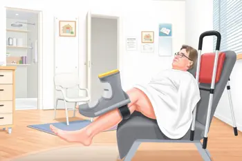
Swelling is expected after surgery and may increase when the leg is positioned downward for prolonged periods. The standard home measures described in the research include rest, ice, compression when specifically permitted, and elevation. Keeping the operated leg elevated is particularly important during the early period, while ice should be applied according to clinical instructions and kept away from a dressing or skin that cannot be inspected. 6
Incision care generally involves keeping the surgical site clean and dry until the treating team provides different instructions. Dressings, casts, and boots should not be modified without guidance. Follow-up assessment is used to evaluate wound healing and tendon integrity. Increasing wound problems, concerning swelling, or a sudden change in function should be communicated to the clinical team rather than managed through unsupervised exercise. 7
Making the home environment safer
Household preparation is a practical part of recovery because crutches and non-weight-bearing restrictions change balance, reach, and the ability to carry objects. Recommended adaptations include a shower chair, clear walking routes, stable elevated surfaces, and non-slip rugs. Frequently used items should be placed within reach, and stairs should be approached only with the method taught by a physical therapist or medical team. 8
Assistive devices can support mobility while the tendon is protected, but they require correct fitting and technique. Crutches or a knee scooter should be used for the prescribed amount of support, and the boot should remain secured during permitted walking. Driving, bathing without protection, and walking without an aid depend on medical clearance, the operated side, vehicle controls, and the patient's ability to respond safely. 9
Physical therapy and home exercises
Physical therapy typically follows the initial immobilization period and progresses from protected range of motion to plantar-flexion strengthening, gait training, balance work, and functional conditioning. Early movement is controlled rather than forceful. Protocols commonly limit dorsiflexion toward neutral at first and avoid forced stretching because excessive lengthening can place stress on the healing tendon. 1
A home exercise program should match the current rehabilitation phase and should not be advanced solely because pain has decreased. Later rehabilitation focuses on rebuilding the gastrocnemius and soleus complex, restoring calf size and strength, and improving walking mechanics. Digital home exercise programs have also been studied as a structured rehabilitation option, but the research emphasizes supervised, progressive rehabilitation rather than unsupervised activity. 10
Longer-term milestones and recovery risks
Many patients require roughly 4 to 6 months to progress from surgery toward full activity, while full tendon strength can take up to 12 months. Low-impact activity may return around the third month in some schedules, but running and court sports are often delayed until approximately 5 to 6 months or longer. High-impact activity is generally considered only after formal strength testing and clinical review. 4
Recovery risks include re-rupture, wound problems, stiffness, persistent weakness, and blood clots associated with prolonged immobilization. Reported re-rupture estimates in the supplied clinical material are approximately 3% to 5% in some surgical data, while DVT prevention may involve aspirin or anticoagulants when prescribed. Medication eligibility and duration depend on personal risk factors, so prophylaxis should be taken only as directed by the treating clinician. 5
Sources
- Cleveland Clinic, Achilles Tendon Rupture: https://my.clevelandclinic.org/health/diseases/22210-achilles-tendon-rupture
- Dr. Nicholas Wessling, Achilles Tendon Repair Recovery Guide: https://www.nicholaswesslingmd.com/post/achilles-tendon-repair-recovery-guide
- MSK Doctors, Recovery Stages After Achilles Tendon Repair: https://mskdoctors.com/insights/unlocking-recovery-what-to-expect-in-each-stage-after-achilles-tendon-repair-surgery
- Mayo Clinic, Achilles Tendon Rupture Diagnosis and Treatment: https://www.mayoclinic.org/diseases-conditions/achilles-tendon-rupture/diagnosis-treatment/drc-20353239
- Balance Foot & Ankle, Achilles Tendon Repair Surgery: https://www.michiganfootdoctors.com/achilles-tendon-repair-surgery-what-to-expect/
- American Academy of Orthopaedic Surgeons, Achilles Tendon Rupture: https://orthoinfo.aaos.org/en/diseases--conditions/achilles-tendon-rupture
- MedlinePlus, Achilles Tendon Repair: https://medlineplus.gov/ency/article/007553.htm
- WebMD, Home Safety After Achilles Tendon Rupture: https://www.webmd.com/fitness-exercise/what-to-know-achilles-tendon-rupture
- Johns Hopkins Medicine, Achilles Tendon Rupture and Assistive Devices: https://www.hopkinsmedicine.org/orthopaedic-surgery/specialty-areas/foot-ankle/conditions/achilles-tendon-rupture
- Wang H. et al., Home-Based Digital Exercise Program After Open Achilles Repair, JMIR mHealth and uHealth: https://www.sciencedirect.com/org/science/article/pii/S2291522226000616
Authored by 24Trendz team