Signs you may have fibrocystic breast disease: A medical analysis of benign breast changes
Understanding the Nature of Fibrocystic Breast Changes
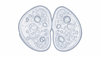
Fibrocystic breast changes, historically referred to as fibrocystic breast disease, represent a common, benign condition characterized by a lumpy or ropelike texture in the breast tissue 6. It is essential to recognize that this condition is not a disease in the traditional sense, but rather a spectrum of normal physiological variations often driven by hormonal fluctuations 6. Medical consensus indicates that these changes are not associated with an increased risk of developing breast cancer 2, 3, 7. The condition is most frequently observed in individuals between the ages of 30 and 50, reflecting the influence of ongoing reproductive hormone activity 5.
The underlying mechanism typically involves the stimulation of milk glands and ducts by estrogen and progesterone during the menstrual cycle 2. When these hormone levels rise, breast tissue may retain fluid, leading to temporary swelling and increased sensitivity 2. As the cycle progresses and hormone levels decline, these symptoms often subside 2. Because this process is tied to internal chemical shifts, the symptoms are often cyclical, manifesting or intensifying in the days leading up to menstruation and improving shortly thereafter 5, 6.
Common Physical Manifestations
The primary indicators of fibrocystic changes include palpable differences in breast texture and discomfort. Individuals often report a generalized lumpiness, particularly in the upper, outer quadrant of the breast near the armpit 2, 6. These areas of thickening often blend into the surrounding breast tissue, though they may feel distinct and nodular to the touch 6. The texture is frequently described as ropey or granular, and these areas can fluctuate in size and consistency over the course of a single month 5, 6.
In addition to physical texture, pain and tenderness are hallmark symptoms for many people 2. This discomfort can range from mild sensations of heaviness to more acute tenderness or burning sensations 2, 7. In some instances, individuals may notice nipple discharge that is green or dark brown in color; this discharge is typically non-bloody and occurs without manual pressure 6. While these symptoms can be persistent, they are generally bilateral, meaning they affect both breasts, although the degree of severity or density may be asymmetric 5, 6.
The Role of Hormonal Factors
Research suggests that prolonged or repeated exposure to fluctuations in estrogen and progesterone is a primary driver of these tissue changes 2, 7. Clinical observations have identified several factors that may increase an individual's susceptibility to experiencing these changes, primarily due to their relationship with estrogen exposure 2, 7. These factors include beginning menstruation at an early age, having a first live birth after the age of 30, or being nulliparous 2, 7.
As individuals approach middle age, the glandular tissue that produces milk begins to transition into softer, fatty tissue, which may alter the way these changes are felt during self-examination 4. It is a notable clinical trend that these symptoms generally subside following the onset of menopause, as the baseline levels of reproductive hormones decrease significantly 2, 7. Understanding this hormonal dependency helps clinicians differentiate between cyclic, benign changes and other conditions that might require different diagnostic approaches 2.
Diagnostic Evaluation and Imaging
Given the variability of breast tissue, clinical assessment is vital to distinguish fibrocystic changes from other types of breast masses. If a patient presents with a concerning lump, clinicians typically initiate a physical examination followed by imaging studies to confirm the benign nature of the tissue 5. For patients under the age of 35, a breast ultrasound is often the primary imaging modality, as it provides clear visualization of fluid-filled cysts without the use of ionizing radiation 5. For those aged 35 or older, a combination of ultrasound and mammography is standard protocol 5.
Imaging results for fibrocystic changes typically reveal dense fibroglandular tissue, potentially accompanied by clustered microcysts or distorted breast parenchyma, while showing an absence of discrete, solid masses that would trigger further concern 5. If imaging remains inconclusive or if a specific lump shows atypical features, a biopsy may be requested to confirm the diagnosis definitively 2, 3. This diagnostic rigor ensures that benign changes are correctly identified, preventing unnecessary anxiety while maintaining a high standard of breast health monitoring 4.
Management and Breast Self-Awareness
Management of fibrocystic breast changes is generally focused on alleviating discomfort rather than curing the condition itself, as it is a natural physiological process 6, 8. Because the condition is common and benign, many individuals find that self-care measures are sufficient to manage daily symptoms 6. Breast self-awareness is highly emphasized; individuals are encouraged to learn the normal baseline feel of their breast tissue, which enables them to identify any new or persistent changes more accurately 4. Any deviation from one's established baseline-such as a new lump that does not resolve or changes in skin appearance-should be reported to a healthcare provider promptly 4, 9.
| Symptom Category | Description |
|---|---|
| Texture | Lumpy, rope-like, or nodular tissue distribution |
| Pain | Cyclical tenderness, heaviness, or burning sensation |
| Timing | Worsens pre-menstruation, improves post-menstruation |
| Discharge | Potential clear, white, or greenish non-bloody fluid |
Distinguishing Benign Conditions
It is important to differentiate fibrocystic changes from other non-malignant breast conditions. While fibrocystic changes involve a broader area of dense or nodular tissue, other conditions such as fibroadenomas manifest as distinct, smooth, mobile, and typically painless masses 7. Other benign occurrences, such as fat necrosis following trauma or the presence of lipomas, present with different clinical features that are distinguishable through palpation and imaging 8. Establishing an accurate clinical picture is essential for patients to distinguish between these varied benign conditions and to manage their health effectively 8.
Healthcare professionals emphasize that while many breast changes are benign, the presence of a new lump should always be professionally evaluated to rule out malignancies 7, 9. By engaging in regular, physician-guided screenings, patients can gain clarity regarding their specific breast tissue profile. This proactive, data-driven approach to breast health ensures that any necessary interventions are provided at the appropriate time while avoiding over-treatment of common, non-problematic tissue variations 4, 8.
Sources
- American Cancer Society: Fibrosis and Simple Cysts in the Breast
- MSD Manual Consumer Version: Fibrocystic Changes of the Breast
- Merck Manual Professional Edition: Breast Masses
- Cedars-Sinai: Fibrocystic Breast Changes
- Osmosis: Fibrocystic breast changes: Clinical Sciences
- Memorial Health System: Fibrocystic breasts
- Franciscan Health: Understanding Cysts And Fibrocystic Breast Disease
- Acibadem International: Understanding Mastopathy Fibrocystic
- National Breast Cancer Foundation: Breast Cyst
Authored by 24Trendz team