Understanding Your Skin Cancer Biopsy Results: A Plain Language Guide
Facts:
- Early detection of skin cancer is crucial for effective treatment.
- Understanding your pathology report empowers patients for informed healthcare decisions.
Demystifying Your Skin Cancer Biopsy Results: What to Expect
Receiving skin cancer biopsy results can be a daunting experience, often filled with anxiety and questions. The medical terminology in a pathology report can be complex, making it difficult to fully grasp what your diagnosis means for your health. This comprehensive guide aims to translate the intricate language of your biopsy results into plain, understandable terms, helping you navigate your diagnosis with clarity and confidence, whether it's a melanoma diagnosis, basal cell carcinoma, or squamous cell carcinoma.
Decoding Your Pathology Report: Essential Components
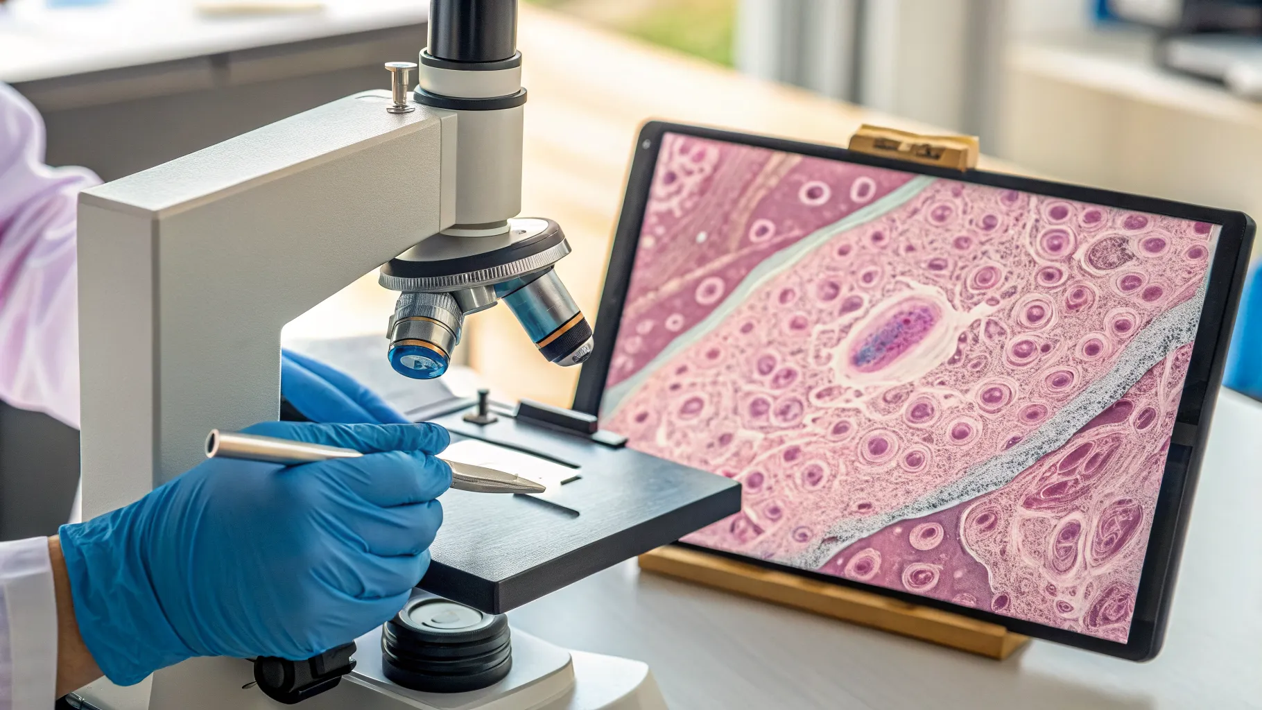
Your pathology report is a vital document that contains the official diagnosis following a skin biopsy. It's compiled by a dermatopathologist, a doctor who specializes in diagnosing skin diseases by looking at tissue samples under a microscope. Understanding the various sections of this report is the first step toward comprehending your skin cancer biopsy results.
Understanding the Biopsy Type and Specimen Description
The report will typically begin by detailing the type of biopsy performed (e.g., shave, punch, excisional, incisional) and a macroscopic (gross) description of the tissue sample. A shave biopsy involves removing only the top layers of skin, while a punch biopsy takes a deeper, circular core. An excisional biopsy removes the entire lesion and a small margin of surrounding healthy skin. The choice of biopsy method can influence what information is available in your skin cancer biopsy results. The specimen description will note the size, color, and any visible abnormalities of the removed tissue.
The Crucial Microscopic Description
This section is the heart of your pathology report. It describes what the dermatopathologist observed when examining the stained tissue slides under the microscope. This can include details about cellular architecture, cell size and shape, nuclear features, mitotic activity, and the presence of inflammatory cells. Terms like "atypia," "dysplasia," "in situ," or "invasive" are commonly found here and are critical to understanding the severity and nature of your skin cancer biopsy results. For instance, "atypia" suggests abnormal cells, while "invasive" indicates the cancer has spread beyond the top layer of skin.
Understanding Specific Diagnoses: Melanoma, Basal Cell, and Squamous Cell Carcinomas
The final diagnosis in your pathology report will identify the specific type of skin cancer, if present. Each type has distinct characteristics and implications for treatment and prognosis.
The Nuances of Melanoma Diagnosis
A melanoma diagnosis is particularly significant because melanoma is the most serious type of skin cancer. The report will specify whether it is melanoma in situ (confined to the epidermis) or invasive melanoma. Key features in the microscopic description for melanoma include atypical melanocytes, pagetoid spread (melanocytes spreading upwards into the epidermis), and mitotic figures. The Breslow thickness (depth of invasion) and Clark level (anatomic level of invasion) are critical measurements for invasive melanoma, directly impacting staging and treatment protocols for your skin cancer biopsy results.
Identifying Basal Cell Carcinoma (BCC)
Basal cell carcinoma is the most common type of skin cancer and is generally slow-growing and rarely metastasizes. Your pathology report will describe nests of basaloid cells with peripheral palisading, retraction artifact, and often a mucinous stroma. Subtypes like nodular, superficial, infiltrative, or morpheaform BCC may also be specified. While BCC is less aggressive, early identification in your skin cancer biopsy results is vital for complete removal and to prevent local tissue destruction.
Diagnosing Squamous Cell Carcinoma (SCC)
Squamous cell carcinoma is the second most common skin cancer and has a higher potential for local recurrence and metastasis compared to BCC. The pathology report will describe atypical keratinocytes with varying degrees of differentiation, keratinization, and intercellular bridges. SCC can be "in situ" (Bowen's disease) or invasive. Features like perineural invasion (cancer cells around nerves) or lymphovascular invasion (cancer cells in lymphatic or blood vessels) are important indicators that affect the prognosis and subsequent management of your skin cancer biopsy results.
The Importance of Margins and Staging in Skin Cancer Biopsy Results
Two critical components of your pathology report that significantly influence treatment decisions are the status of the surgical margins and, for more aggressive cancers like melanoma, the staging information.
Assessing Surgical Margins
Surgical margins refer to the edges of the tissue removed during the biopsy. A "clear margin" or "negative margin" means that no cancer cells were found at the edges of the excised tissue, suggesting the entire tumor was removed. A "positive margin" indicates that cancer cells are present at the edge, meaning some cancer may have been left behind. In such cases, further surgery may be recommended to achieve clear margins. Understanding margin status is crucial for guiding follow-up treatment after receiving your skin cancer biopsy results.
Understanding Tumor Staging
For invasive melanomas and aggressive squamous cell carcinomas, the pathology report will contribute to the overall tumor staging. Staging combines information from the biopsy (like Breslow thickness for melanoma), clinical examination, and sometimes imaging studies to determine the extent of the cancer. Staging helps classify the cancer into numerical stages (e.g., Stage I, II, III, IV), which guides prognosis and treatment plans. This comprehensive assessment ensures that your treatment strategy is tailored to the specific nature and spread of your melanoma diagnosis or other advanced skin cancer.
Empowering Yourself: Next Steps and Actionable Advice
Receiving and interpreting your skin cancer biopsy results is just the beginning. Proactive engagement with your healthcare team is essential for optimal outcomes.
- Ask Questions: Come prepared to your follow-up appointment with a list of questions. Don't hesitate to ask your dermatologist to explain any terms in your pathology report that you don't understand. Inquire about the specific type of cancer, its invasiveness, margin status, and what this means for your overall prognosis.
- Seek a Second Opinion: For complex or ambiguous skin cancer biopsy results, or for a melanoma diagnosis, considering a second opinion from another dermatopathologist or a specialized cancer center can provide peace of mind and confirm the diagnosis and treatment plan.
- Understand Your Treatment Options: Based on your pathology report, your doctor will discuss various treatment modalities. These could range from further excisions, Mohs surgery for basal cell carcinoma and squamous cell carcinoma, radiation therapy, or systemic therapies for advanced melanoma. Ensure you understand the benefits, risks, and recovery associated with each option.
- Regular Follow-ups and Self-Exams: Regardless of your specific skin cancer biopsy results, regular follow-up appointments with your dermatologist are crucial for monitoring for new lesions or recurrences. Performing regular self-skin exams can also help detect changes early.
Frequently Asked Questions
What does 'in situ' mean in a skin cancer biopsy result?
'In situ' means the cancer cells are confined to the very top layer of the skin (epidermis) and have not invaded deeper tissues. For example, 'melanoma in situ' or 'squamous cell carcinoma in situ' (Bowen's disease) are generally considered early-stage cancers with a very high cure rate, typically with simple excision.
How long until I get my pathology report?
The time it takes to receive your skin cancer biopsy results can vary, but it typically ranges from a few days to two weeks. Factors influencing this include the complexity of the case, the specific tests required by the pathologist, and the workload of the lab. Your doctor's office will usually contact you when the report is ready to discuss the findings.
Is a positive margin always bad after a melanoma diagnosis?
A positive margin in a melanoma diagnosis means that cancer cells were present at the edge of the removed tissue, indicating that some melanoma may still be in your body. While it requires further intervention, it doesn't automatically mean a poor prognosis. Often, a subsequent wider excision can successfully remove any remaining cancer cells, achieving clear margins and preventing recurrence.
Can basal cell carcinoma spread to other parts of the body?
While basal cell carcinoma (BCC) is typically a slow-growing cancer that rarely metastasizes (spreads to distant sites), it can be locally destructive if left untreated. In extremely rare cases, aggressive BCCs can spread, but this is highly uncommon. Early detection and treatment based on your skin cancer biopsy results are key to preventing local tissue damage and recurrence.
References
- American Academy of Dermatology Association. (n.d.). Skin Cancer: Types, Causes, Symptoms, and Treatment.
- National Cancer Institute. (n.d.). What You Need To Know About Skin Cancer.
- Mayo Clinic. (n.d.). Skin biopsy: What you can expect.
Authored by 24Trendz team