Understanding Common Skin Cancer Indicators and Red Flags
Facts:
- Early detection of skin cancer dramatically improves treatment outcomes.
- Globally, incidence of melanoma red flags is increasing for all age groups.
Recognizing the Early Signs of Skin Cancer
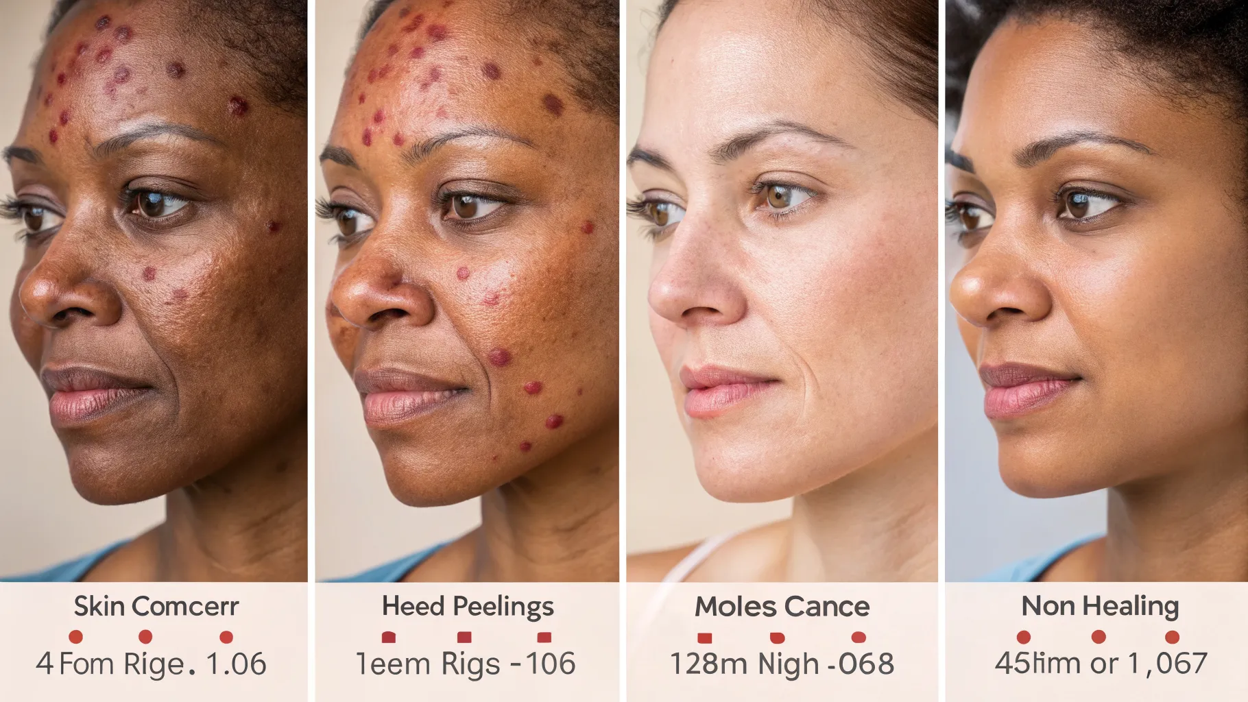
Identifying common skin cancer indicators early is paramount for effective treatment and improved prognosis. Many people overlook subtle changes in their skin, mistaking them for harmless blemishes, but understanding the red flags can be life-saving. This comprehensive guide will delve into the various types of skin cancers and their distinct warning signs, empowering individuals to be proactive about their dermatological health and seek timely medical advice for any suspicious lesions or changes on their skin.
Melanoma: The ABCDEs of Early Detection
Melanoma is often considered the most serious type of skin cancer due to its potential to spread rapidly if not caught early. Recognizing melanoma red flags involves a systematic approach known as the ABCDE rule. This mnemonic helps individuals identify characteristics of suspicious moles or lesions that warrant medical evaluation.
Asymmetry: One Half Doesn't Match the Other
Normal moles are typically symmetrical. If you draw a line through the middle of a mole and the two halves do not match, it is a sign of asymmetry. This lack of symmetry is one of the crucial common skin cancer indicators and a significant red flag for potential melanoma. Consistently monitoring for changes in symmetry is vital.
Border: Irregular, Jagged, Notched, or Blurry Edges
The borders of benign moles are usually smooth and well-defined. In contrast, melanoma red flags often include borders that are irregular, ragged, notched, or blurred. These ill-defined edges suggest uncontrolled growth patterns characteristic of cancerous cells. Any mole displaying such characteristics should be examined by a dermatologist to rule out skin cancer.
Color: Uneven Coloration, Shades of Black, Brown, and Tan
A mole with uneven coloration or varying shades of black, brown, and tan within the same lesion is a significant warning sign. The presence of red, white, or blue hues can also indicate advanced melanoma. Uniform color is typical for harmless moles, while diverse pigmentation points towards common skin cancer indicators that require immediate attention.
Diameter: Larger Than 6 Millimeters (About the Size of a Pencil Eraser)
While some melanomas can be smaller, a diameter greater than 6 millimeters (approximately the size of a pencil eraser) is a common criterion for suspicion. However, it's crucial to remember that some melanomas can be smaller, so size alone isn't the only factor. Monitoring for growing moles, regardless of initial size, is an important part of identifying melanoma red flags.
Evolving: Any Change in Size, Shape, Color, or New Symptoms
The most critical factor in the ABCDE rule is 'E' for Evolving. Any change in a mole's size, shape, color, elevation, or the development of new symptoms such as bleeding, itching, or tenderness, is a significant skin cancer indicator. Constant vigilance for evolution of existing moles or the appearance of new, unusual spots is essential for early diagnosis of melanoma red flags.
Basal Cell Carcinoma: The Most Common Skin Cancer
Basal cell carcinoma signs represent the most frequent type of skin cancer, often appearing as a slow-growing lesion that rarely spreads to other parts of the body. Despite its generally less aggressive nature compared to melanoma, it can cause significant local tissue damage if left untreated. Understanding these signs is crucial for early intervention.
Pearly or Waxy Bumps
One of the classic basal cell carcinoma signs is a pearly or waxy bump, often with a translucent quality. These bumps may be skin-colored, pink, or red and might have visible blood vessels (telangiectasias) on the surface. They typically appear on sun-exposed areas like the face, ears, neck, or hands. People often mistake these for a harmless pimple that doesn't go away.
Flat, Flesh-Colored or Brown Scar-Like Lesions
Another presentation of basal cell carcinoma signs can be a flat, flesh-colored, or brownish lesion that resembles a scar. These lesions can be firm to the touch and might have slightly raised borders. They often appear on the back or chest and can be easily overlooked as an insignificant mark on the skin, underscoring the need for careful self-examination.
Open Sores That Bleed, Ooze, or Crust and Don't Heal
Any sore that bleeds, oozes, or crusts and remains open for several weeks is a definite red flag. These non-healing sores are a strong indicator of basal cell carcinoma signs, even if they seem to heal temporarily before reopening. Persistent ulceration demands immediate medical attention as it is a highly concerning common skin cancer indicator.
Squamous Cell Carcinoma: Growing and Spreading Potential
Squamous cell carcinoma symptoms are the second most common type of skin cancer, originating in the flat cells that make up the outer part of the epidermis. While more likely to spread than basal cell carcinoma, it is usually curable if detected and treated early. These cancers often develop on sun-exposed skin, such as the face, ears, neck, lips, and back of the hands.
Firm, Red Nodules
One common presentation of squamous cell carcinoma symptoms is a firm, red nodule. These nodules can be tender to the touch and may grow rapidly. Unlike a simple pimple, these lesions persist and often become more pronounced over time. They are often found on areas heavily exposed to the sun, making regular checks of these regions essential.
Scaly, Crusted Patches
Scaly, crusted patches are another significant indicator of squamous cell carcinoma symptoms. These patches may resemble warts or open sores and can bleed easily if bumped or scraped. They can feel rough and may be painful or itchy. This type of lesion highlights the importance of not dismissing persistent rough skin patches, especially on sun-damaged skin, as merely dry skin.
Precancerous Lesions: Actinic Keratoses
Beyond active cancers, understanding precancerous lesions is vital for preventing the development of full-blown skin cancer. Actinic keratoses (AKs) are rough, scaly patches that develop on skin heavily damaged by UV radiation. They are considered early warning signs because they have the potential to progress into squamous cell carcinoma.
Rough, Dry, or Scaly Patches
Actinic keratoses typically appear as rough, dry, or scaly patches, often feeling like sandpaper. They vary in color, from skin-toned to reddish-brown or dark pink. These lesions are generally small, often less than 1 centimeter in diameter, and can be more easily felt than seen. Identifying these precancerous lesions early allows for simple, non-invasive treatments to prevent progression to squamous cell carcinoma symptoms.
Often Found on Sun-Exposed Areas
These precancerous lesions are almost exclusively found on areas of the skin that receive significant sun exposure, such as the face, scalp, lips, and the back of the hands and forearms. People with fair skin, a history of extensive sun exposure, or weakened immune systems are particularly susceptible. Regular self-examinations and professional skin checks are critical for identifying and treating these early indicators of potential skin cancer.
Other Warning Signs and When to See a Doctor
Beyond the specific characteristics of melanoma, basal cell carcinoma, and squamous cell carcinoma, there are general common skin cancer indicators and red flags that should prompt a visit to a dermatologist. Any new or changing lesion, especially one that persists for more than a few weeks, warrants professional evaluation.
New Growth or Spot That Looks Different from Others
The 'ugly duckling' sign is a useful concept: if one mole or spot looks distinctly different from all the others on your body, it should be examined. This could be a new, unusually shaped growth or an existing lesion that has started to change significantly. This distinctiveness can be a powerful melanoma red flag, even if it doesn't perfectly fit the ABCDE criteria.
A Sore That Doesn't Heal, or Recurrent Bleeding from a Spot
Any sore or wound on the skin that does not heal within a few weeks, or repeatedly bleeds, crusts, or oozes, is a serious common skin cancer indicator. This is particularly true for lesions that seem to heal partially and then reopen. Persistent bleeding from a mole or skin lesion, even without trauma, should be reported to a doctor immediately as it can be a sign of advanced basal cell carcinoma signs or other forms of skin cancer.
Itching, Tenderness, or Pain in a Mole or Lesion
While many moles are harmless, the development of new symptoms such as itching, tenderness, or pain in an existing mole or a newly formed lesion can be a red flag. These sensations are not typical of benign moles and can sometimes indicate an underlying malignancy, including various forms of skin cancer. Any persistent discomfort associated with a skin spot should be evaluated by a healthcare professional.
Proactive Steps for Skin Health and Early Detection
Being proactive about skin health is the best defense against skin cancer. This involves a combination of preventative measures and diligent self-monitoring. Integrating these practices into your routine can significantly reduce your risk and improve the likelihood of early detection if any issues arise.
- Perform Monthly Self-Skin Exams: Regularly examine your entire body for any new moles, changes in existing moles, or unusual spots. Pay close attention to areas not easily seen, using a mirror or asking a partner for help. Knowing your skin's normal appearance makes it easier to spot common skin cancer indicators.
- Protect Your Skin from UV Radiation: Limit direct sun exposure, especially during peak hours (10 AM to 4 PM). Use broad-spectrum sunscreen with an SPF of 30 or higher daily, even on cloudy days. Wear protective clothing, wide-brimmed hats, and UV-blocking sunglasses to shield your skin from harmful rays that contribute to melanoma red flags.
- Schedule Annual Professional Skin Checks: Even with diligent self-exams, a dermatologist can identify suspicious lesions that you might miss. Annual full-body skin examinations by a professional are crucial, especially for individuals with a history of extensive sun exposure, a high number of moles, or a family history of skin cancer.
- Avoid Tanning Beds: Tanning beds emit harmful UV radiation that significantly increases the risk of all types of skin cancer, including melanoma. The artificial UV exposure contributes to premature skin aging and elevates the risk of developing basal cell carcinoma signs and squamous cell carcinoma symptoms.
- Know Your Family History: A family history of skin cancer, particularly melanoma, increases your personal risk. Be sure to inform your dermatologist about any family history so they can tailor screening recommendations and provide more personalized advice regarding precancerous lesions and overall skin health.
Frequently Asked Questions
What are the most common skin cancer indicators?
The most common indicators include changes in moles (ABCDEs for melanoma), pearly or waxy bumps, flat scar-like lesions, and non-healing sores (basal cell carcinoma), or firm red nodules and scaly patches (squamous cell carcinoma). Any persistent new or changing lesion should be evaluated.
How often should I check for melanoma red flags?
It is recommended to perform a self-skin exam monthly. Additionally, annual professional skin checks with a dermatologist are crucial, especially if you have risk factors such as numerous moles, fair skin, or a family history of skin cancer.
Are precancerous lesions always a sign of cancer?
No, precancerous lesions like actinic keratoses are not cancer themselves, but they have the potential to develop into squamous cell carcinoma over time. Early identification and treatment can prevent their progression to full-blown skin cancer, highlighting the importance of early intervention.
References
- American Academy of Dermatology Association. Skin Cancer.
- Skin Cancer Foundation. Early Detection and Self-Exams.
- National Cancer Institute. Basal Cell Carcinoma and Squamous Cell Carcinoma.
- Mayo Clinic. Melanoma Diagnosis and Treatment.
- Centers for Disease Control and Prevention. Sun Protection.
Authored by 24Trendz team